A tendon problem can develop gradually from repeated loading or happen suddenly when a tendon partially or completely tears, and those injuries do not follow the same treatment path. You will learn how to identify the likely problem, protect it during the first few days, understand the assessment and scans, and judge treatment and return-to-sport decisions.
Key takeaways
- Pain with resisted movement often points to a tendon problem.
- Use relative rest, ice, compression, and elevation during the first 24 to 72 hours.
- Ultrasound shows many tendon tears; MRI helps assess deeper or complex injuries.
- Return to sport only after strength, movement, and sport-specific loading recover.
Is the problem a tendon injury, muscle strain, ligament injury, joint injury, or stress fracture?
Pain location, how the injury happened, and what triggers it usually separate a tendon injury from a muscle, ligament, joint, or bone problem.
| Pattern | Typical clues |
|---|---|
| Tendon injury or tendinopathy | Pain sits along a tendon and builds with repeated loading, then eases with rest. Achilles tendinopathy affects the back of the ankle; patellar tendinopathy sits below the kneecap; tennis elbow affects the outer elbow; rotator-cuff tendinopathy causes pain around the shoulder; de Quervain tenosynovitis affects the thumb side of the wrist. |
| Muscle strain | Pain follows a forceful contraction or sudden stretch and may include bruising, weakness, or tenderness within the muscle. |
| Ligament injury | Twisting, a joint displacement, or a sudden change of direction causes pain beside the joint, instability, and swelling. |
| Joint injury | Deep swelling, stiffness, catching, locking, or painful movement points toward cartilage or other structures inside the joint. |
| Stress fracture | Focal tenderness directly over bone worsens with impact and may progress to pain during ordinary walking. |
Tendinopathy reduces a tendon’s load tolerance over time without requiring a discrete tear. Achilles tendinopathy diagnosis clues include pain at the back of the ankle during running or heel raises, morning stiffness, and tenderness along the tendon.
An acute tendon tear follows a sudden force and can cause a pop, immediate weakness, a gap, deformity, or inability to push off; partial and complete tears need prompt assessment.
What should you do during the first 24 to 72 hours?
During the first 72 hours after a tendon injury, stop the movement that triggered pain and protect the area from impact, heavy loading, and sudden stretching. Use a brace, crutches, or protected walking if normal movement causes pain, weakness, or instability.
For ice for tendon injury, wrap a cold pack in cloth and apply it for about 15 to 20 minutes. Check the skin between applications; stop if it becomes white, numb, painful, or unusually cold. Use light compression only if it does not cause numbness, tingling, colour change, or increasing pain.
Compression and elevation work together: raise the injured area above heart level when swelling is present.
Do not repeatedly test strength, force a painful stretch, or use aggressive massage when a tear is possible. These actions can increase bleeding, swelling, or tissue damage.
Seek prompt assessment after any of these warning signs:
- A pop or visible deformity
- Sudden loss of strength
- Inability to bear weight or push off
- Inability to perform a heel raise
- Rapidly increasing swelling
- Numbness
- A hot, red joint with fever
Treat a suspected tendon rupture as urgent, especially when you cannot actively move the joint normally. Do not drive yourself if you cannot safely control the vehicle.
What happens during the examination, and which scan is useful?
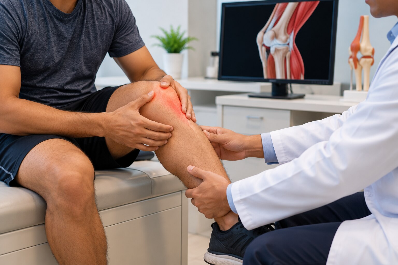
The visit starts with a history and a tendon injury examination. The clinician asks which activity caused the problem, how quickly symptoms appeared, whether you have had previous injuries, what your work and sport demand, and whether your training load recently changed.
Examination includes looking for swelling or contour changes, checking range of motion, testing resisted movement and strength, and comparing the painful side with the uninjured side.
| Scan | What it shows | What it cannot establish |
|---|---|---|
| X-ray | Bone alignment, fractures, and sometimes tendon calcification | Most tendon injuries directly; these are the key X-ray tendon injury limitations |
| Ultrasound | Tendon structure, movement, inflammation, and many partial or complete tears in real time | Every deep or complex injury; image quality depends on the operator |
| MRI | Detailed views of the tendon, muscle, bone, joint, and surrounding tissues | Whether an abnormal finding is the pain source by itself |
An ultrasound tendon tear assessment is useful when the clinician needs to watch the tendon move. An MRI tendon injury assessment helps map extensive damage or nearby joint and muscle problems.
A scan must match weakness, function, injury history, and examination findings. Tendon abnormalities can exist without pain, so an abnormal image is not an automatic reason for surgery. A normal-looking scan also cannot replace a careful examination when strength or function has suddenly changed.
How is the treatment plan chosen for tendinopathy, a partial tear, or a rupture?
The plan depends on whether the tendon is overloaded, partly torn, or fully ruptured, not on the scan alone. Clinicians weigh the tendon involved, injury severity and duration, age, sport and work demands, tendon quality, rupture status, diabetes, smoking, muscle atrophy, and your treatment goal.
| Option | What it means | When it applies |
|---|---|---|
| Observation | Modify painful activity and monitor strength and function | Mild symptoms, preserved function, or improvement with load reduction |
| Tendon rehabilitation | Restore capacity through progressive tendon loading, strength, and task-specific work | Most tendinopathy and many stable partial tears |
| Injection | Temporary symptom control, not tendon reconstruction | Selected cases when pain blocks rehabilitation |
| Procedure | Repair or reconstruct damaged tendon | Complete rupture, major weakness, failed rehabilitation, or high functional demands |
For chronic Achilles tendinopathy, try progressive tendon loading at least three times weekly for a minimum of 12 weeks before calling rehabilitation ineffective. Heavy slow resistance and eccentric exercise can both work; tolerable progression matters more than one special exercise. Rest alone often lowers pain without restoring the tendon’s capacity for running, jumping, or work.
A corticosteroid tendon injection can provide short-term relief in selected conditions, but it does not repair tendon tissue; corticosteroid tendon injection risks include weakening with repeated or intratendinous exposure. PRP tendon injection evidence remains inconsistent, so PRP is an option, not guaranteed regeneration.
Complete Achilles rupture can suit surgical repair or functional nonoperative rehabilitation. A traumatic full-thickness rotator-cuff tear with major weakness makes surgery a more serious consideration, especially when function continues to decline.
How long will recovery take, and when is it safe to return to sport or heavy work?
Recovery is measured in months for a repaired or ruptured tendon, not weeks. A calendar date or improved everyday pain is not enough: pain can settle while the tendon still lacks the strength and load tolerance needed for sport or heavy work.
Tendon injury recovery time depends on the tendon, tear size, tissue quality, and your demands. Tendon repair rehabilitation usually starts with protection or immobilization, followed by controlled movement, strengthening, and later running, jumping, throwing, or pivoting drills. Achilles rupture recovery follows this staged pattern.
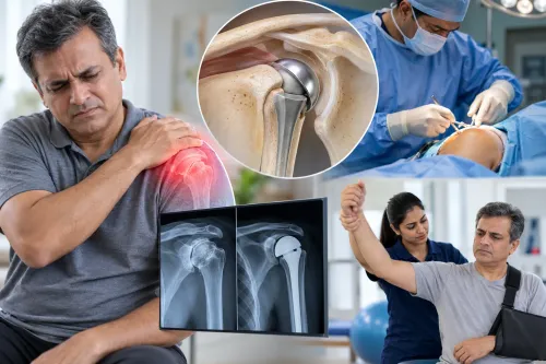
Rotator cuff repair recovery also progresses in stages: protected motion in a sling, active motion, then strengthening while the repair regains biological strength. Feeling better does not mean the repair is ready for lifting.
Before return to sport after tendon injury or heavy work, check that you have:
- Full or near-full range of motion compared with the uninjured side
- Near-symmetric strength and endurance
- Sport- or job-specific loading without pain or a next-day flare
- Clearance from the treating clinician or physiotherapist
Larger tears, poor tendon quality, older age, diabetes, smoking, and muscle atrophy can slow healing and increase complication or repair-failure risk. A normal-looking scan alone cannot confirm readiness.
When diagnosis or treatment choice remains unclear, Dr Gaurav Gupta can match examination findings and imaging with rehabilitation progress and the demands of your sport or work.
Related services
Tendon Injury Treatment Tendon injuries can cause pain, swelling, and restricted movement, affecting daily activities. Dr. View service → |
Sports Injury Treatment Dr. Gaurav Gupta offers advanced diagnosis and treatment for sports-related injuries, including ligament tears, muscle strains, fractures, and joint... View service → |
Frequently asked questions
How can you tell a tendon injury from a muscle, ligament, joint, or bone injury?
Pain location, the injury mechanism, swelling, bruising, and the movement that triggers pain help distinguish these conditions. Tendon pain often worsens when you contract or stretch the attached muscle, while a ligament injury follows a joint-twisting event and a stress fracture causes focused bone tenderness that worsens with impact.
What should you do during the first 24 to 72 hours after a sports or overuse injury?
Stop the activity that causes pain, use relative rest, apply a covered ice pack for 15 to 20 minutes at a time, add comfortable compression, and elevate the limb. Avoid aggressive stretching, deep massage, heat, and returning to sport while pain or swelling is increasing. Seek urgent care for a visible deformity, a sudden pop with weakness, inability to bear weight, numbness, or rapidly increasing swelling.
What happens during a tendon injury examination, and which scan is useful?
The clinician checks the painful area, swelling, bruising, range of motion, strength, tendon continuity, and nearby joints. Ultrasound can show tendon thickening, inflammation, and many partial or complete tears in real time. MRI gives a wider view and helps assess deep tendons, complex tears, bone stress injury, and associated muscle or ligament damage.
How is treatment chosen for tendinopathy, a partial tear, or a rupture?
Treatment depends on the tendon involved, tear size, loss of strength, activity demands, and examination or scan findings. Tendinopathy often uses load modification and progressive strengthening. A partial tear may require bracing, protected weight-bearing, and rehabilitation. A complete rupture with major functional loss can require prompt surgical assessment, followed by structured rehabilitation.
How long does tendon injury recovery take, and when can you return to sport or heavy work?
Recovery ranges from several weeks for a mild overload problem to several months for a substantial tear or surgery. Return when daily activity is pain-free, swelling has settled, movement is near the uninjured side, strength is restored, and you can complete progressive sport- or work-specific drills without symptoms. A clinician or physiotherapist should clear high-load activity after a rupture or significant tear.